
Visualizing Breath
Wearing face coverings is a simple way to reduce the spread of airborne illnesses. The invisible nature of human breath, however, makes it hard to directly observe the effectiveness of different mask materials and designs.
In collaboration with the Department of Ophthalmology at Stanford and science educators at the Exploratorium, I built Schlieren imaging systems to visualize breath and bring awareness to best practices for face coverings in clinical and social settings.
Ophthalmology Clinics
In June 2020, I partnered with ophthalmologists in the Stanford School of Medicine to develop best practices for using face coverings in ophthalmology clinics. Our concern was that the redirected plume of breath from a face mask could contaminate the multi-dose eye drop bottles that are reused from patient to patient, which could increase the risk of
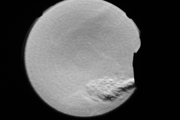
Visualization of breath plumes in profile with a Schlieren imaging system
Seeing Your Breath: Using Science to Choose the Proper Mask
To build the Schlieren system for Stanford, I needed to find a large concave mirror. I had previously done educational outreach events at the Exploratorium and remembered their